Many creatures on Earth are known for their ability to emit a visible glow, yet humans have rarely been considered among them. However, recent scientific investigations suggest this assumption may not be entirely accurate. Since as far back as 1923, studies have indicated that humans emit a faint luminescence, though it is too weak for the naked eye to detect. This subtle glow, present from conception until death, may hold secrets about our internal processes.
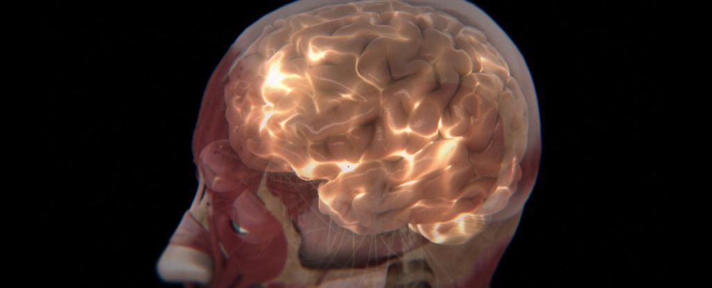
In a groundbreaking study, a team led by biologist Hayley Casey from Algoma University in Canada has focused on the brain’s weak glow. By meticulously recording the faint emissions from the human brain, they discovered that these emissions vary with brain activity. This finding paves the way for a novel technique, termed photoencephalography, which could potentially assess brain health by reading these ‘biophotons’.
Understanding Ultraweak Photon Emissions
The concept of ultraweak photon emissions (UPEs) is not entirely new. Everything in the universe above absolute zero emits some form of radiation, including humans. While thermal radiation is well understood, UPEs are distinct, occurring in near-visible to visible wavelengths. These emissions result from electrons releasing photons as they lose energy—a natural by-product of metabolism.
Casey and her colleagues aimed to differentiate brain UPEs from other background radiation and to determine if these emissions correlate with different brain activity levels. Their study involved placing participants in a dark room, using electroencephalography (EEG) caps to monitor brain activity, and employing photomultiplier tubes to detect light emissions. These tubes are highly sensitive and capable of detecting even the faintest light.
“As the first proof-of-concept demonstration that ultraweak photon emissions (UPEs) from human brains can serve as readouts to track functional states, we measured and characterized photon counts over the heads of participants while they rested or engaged in an auditory perception task,” the researchers noted in their paper.
Linking Brain Activity with Light Emissions
The study’s results were compelling. Not only were UPEs measurable from outside the participants’ heads, but there was also a clear correlation between UPE output and EEG-detected brain activity. This correlation suggests that UPEs could potentially be used to monitor brain states, providing a non-invasive method to assess neurological health.
Future research could explore how neuroanatomy influences UPE output and whether different activities produce distinct UPE patterns. Additionally, researchers are curious if individuals have unique UPE ‘fingerprints’ that could serve as baselines for identifying abnormal activity.
“We view the current results as a proof-of-concept demonstration that patterns of human-brain-derived UPE signals can be discriminated from background light signals in darkened settings despite very low relative signal intensity,” the researchers emphasized.
Implications and Future Directions
The implications of this research are significant. If further studies confirm these findings, photoencephalography could become a valuable tool in neuroscience and medicine. It might help in diagnosing and monitoring various brain disorders, offering a new dimension to understanding brain function.
Moreover, the potential to enhance UPE signal features using selective filters and amplifiers could lead to breakthroughs in distinguishing between healthy and diseased brain states. As the research progresses, it may also shed light on the fundamental processes of human metabolism and energy emission.
The study, published in Current Biology, marks a promising step toward unlocking the mysteries of the human brain’s luminescence. As scientists continue to explore this phenomenon, the hope is to develop practical applications that could revolutionize our approach to brain health.